 Diagnose
Diagnostic Intake
A pathologist is
already asking
these questions.
Answer three questions about your report. We'll match you to the right subspecialist and show you exactly what your slide should contain.
Specimen processing: Step 1 of 3
What type of report are you holding?
Select the specimen category that matches your lab report header.

Turnaround
24h
routine biopsy reads
Currently reviewing

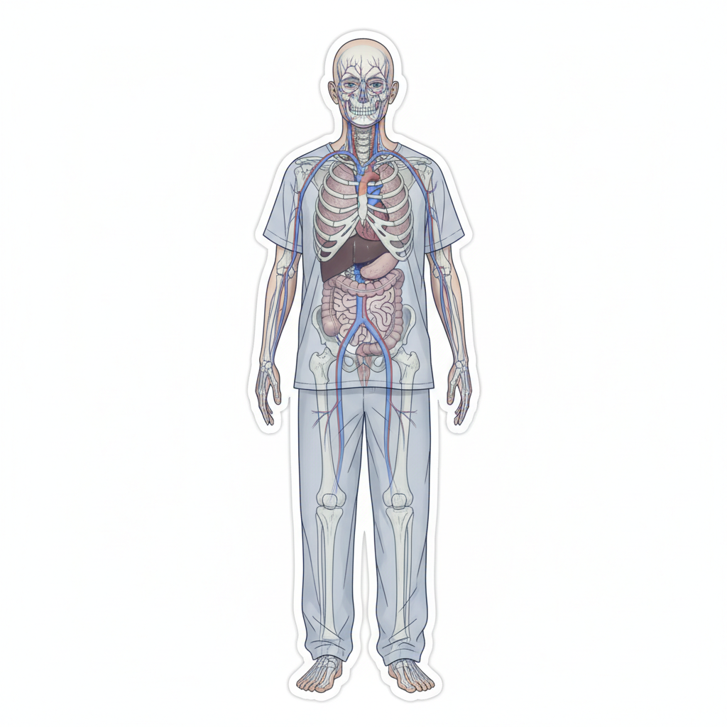

active cases today
Diagnostic Reference
Conditions & Diagnostic Checklists
Each entry reflects how our pathologists approach the slide — signs observed, stains ordered, and what your results mean in plain language. Conditions are ordered from common to complex.
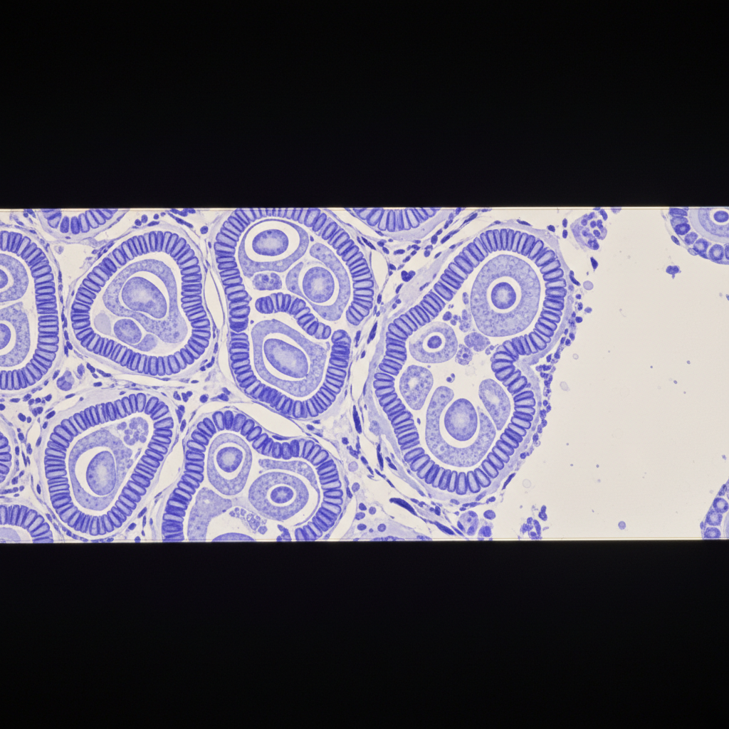
Basal Cell Carcinoma
The most frequently diagnosed skin cancer. Highly curable when caught early, arising from basal cells in the epidermis.
Signs We Look For
Palisading nuclei at periphery
Columnar cells arranged at tumor border — pathognomonic feature
Retraction artifact
Cleft between tumor nests and stroma in formalin-fixed specimens
Mucinous stroma
Pale blue myxoid matrix surrounding nests
Mitotic figures
Low to moderate; high count suggests more aggressive subtype
Staining Protocols
BerEP4 IHC
Positive in BCC; distinguishes from SCC and other basaloid tumors
Mohs mapping
Required for high-risk anatomic sites (periorbital, nasal tip, ear)
Typical Turnaround
24–48 hours
Plain Language Explanation
"Your biopsy shows cancer cells growing from the deepest layer of your skin. This type rarely spreads to other parts of the body. The pathologist checks the edges of the sample to see if all the cancer cells were removed."
Does your current report include these markers?
Check each item against your current pathology report.
Missing items in your report? Our pathologists can complete a full diagnostic workup.
Request a complete workup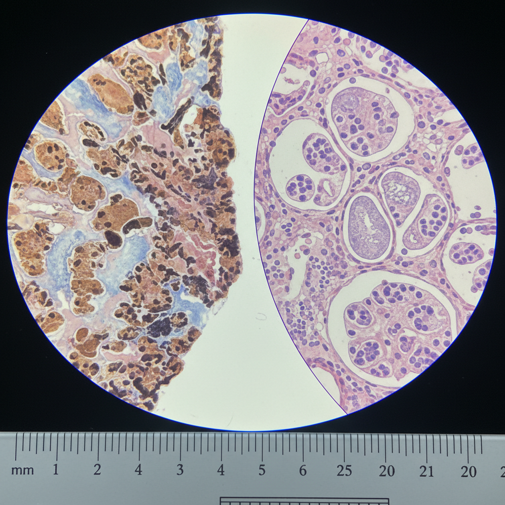
Melanoma, Superficial Spreading
The most common melanoma subtype. Prognosis depends critically on Breslow thickness and ulceration status — both must be in your report.
Signs We Look For
Pagetoid spread
Melanocytes scattered throughout epidermis above basal layer
Breslow thickness
Millimeter measurement from granular layer to deepest tumor cell — most critical prognostic factor
Clark level
Anatomic depth: I (epidermis) through V (subcutis)
Ulceration
Full-thickness epidermal loss over tumor — upstages T category
Mitotic rate
Mitoses/mm² in dermis — required for T1 staging
Staining Protocols
SOX10 + MART-1 panel
Confirm melanocytic lineage; SOX10 more sensitive for spindle cell variants
BRAF V600E testing
Required for metastatic disease; guides targeted therapy eligibility
Sentinel lymph node mapping
Recommended when Breslow > 0.8mm or ulceration present
Typical Turnaround
48–72 hours (FISH/NGS adds 5–7 days)
Plain Language Explanation
"Melanoma is a skin cancer that starts in the pigment-producing cells. The most important numbers in your report are the thickness (Breslow depth) and whether the surface is broken (ulceration). These two facts determine your treatment plan more than anything else."
Does your current report include these markers?
Check each item against your current pathology report.
Missing items in your report? Our pathologists can complete a full diagnostic workup.
Request a complete workupOur Pathologists
Board-Certified. Fellowship-Trained.
Reading your slide today.
Every report is signed by a physician with subspecialty training in the tissue type submitted. No generalists, no outsourced reads.
18+
Years Average Experience
Per board-certified pathologist on staff
24h
Routine Turnaround
Most biopsy reports completed next business day
6
Subspecialty Panels
Dermato, Heme, GI, Breast, Neuro, Gynecologic
4h
Frozen Section
Intraoperative consultation available

Dr. Margaret Holloway, MD
Dermatopathology · Mohs Surgery
Memorial Sloan Kettering, 2009
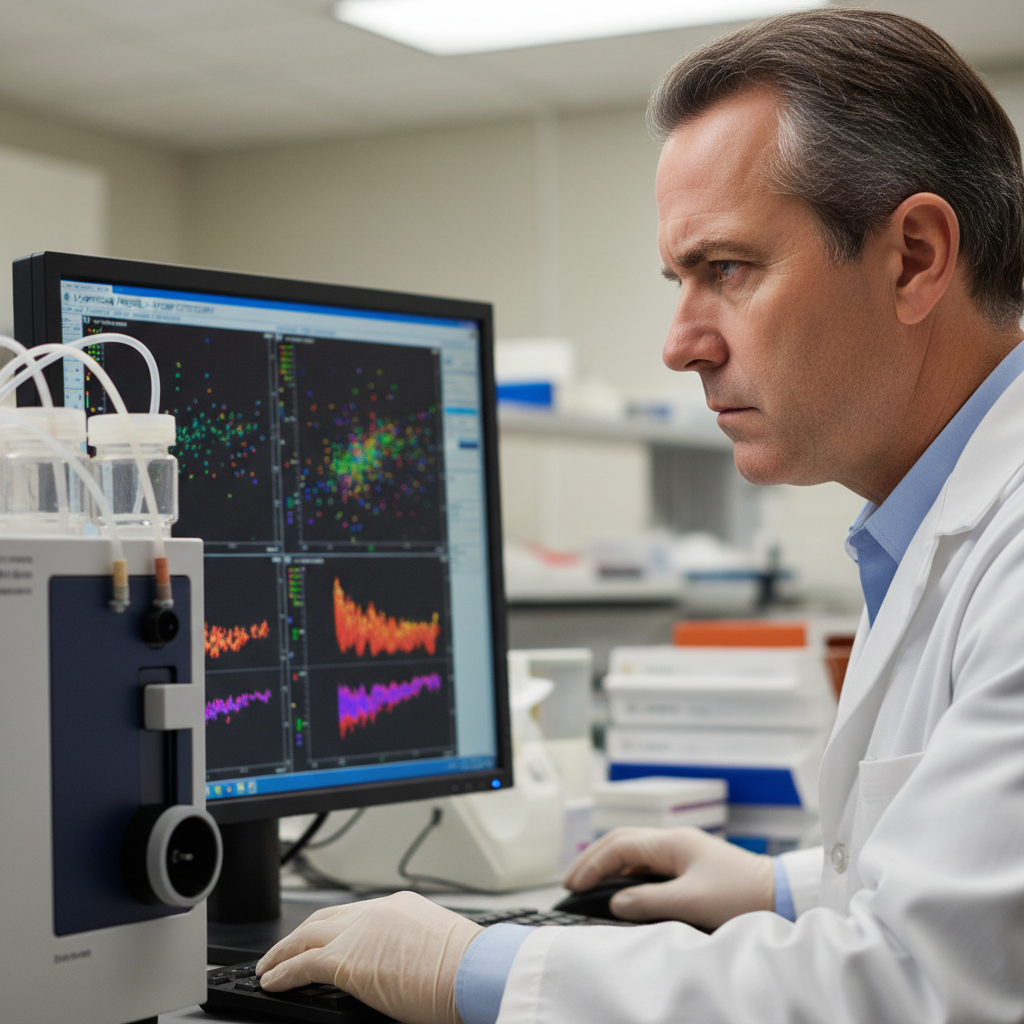
Dr. James Okonkwo, MD PhD
Lymphoma Subtyping · Flow Cytometry
Mayo Clinic Hematopathology, 2011

Dr. Priya Venkataraman, MD
GI Oncology · Inflammatory Bowel
Johns Hopkins GI Pathology, 2013
Real Outcomes
From the patients,
physicians, and partners who trust us.
Three distinct users. One shared expectation: accurate, fast, and clearly communicated pathology.
"I referred a lymphoma case on a Tuesday afternoon expecting a week-long wait. Diagnose returned a complete FISH panel and cell-of-origin classification by Thursday morning. My oncology colleagues were able to start R-CHOP within the week."

Dr. Rebecca Hartmann, MD
Oncologist · Pacific Northwest Cancer Associates, Portland OR
"My dermatologist sent a biopsy report saying 'atypical melanocytic proliferation' and I had no idea if I should be terrified. The checklist on this site explained exactly what markers were missing. When I asked my dermatologist about Breslow depth, she ordered a re-read. That re-read changed my treatment."

Natasha Obumseli
Patient, Stage IB Melanoma Survivor · Austin, Texas
"We send all our GI biopsies to Diagnose for second-opinion reads before discussing results with patients. The turnaround is predictable, the reports are granular, and the pathologists pick up the phone. That last part matters more than I expected."

Dr. Marcus Chen, MD
Gastroenterologist · Chicago Digestive Health Group, Chicago IL
"As a clinic manager, I was evaluating three pathology partners for our send-out work. Diagnose was the only one with a public-facing turnaround SLA and a real person who answered my questions about CLIA certification within the hour."

Sandra Mbeki
Laboratory Operations Manager · Cascade Regional Medical Group, Seattle WA
Book Now
Someone is already reading your slide.
Let's make it official.
Choose how you'd like to proceed. Both pathways are reviewed by a board-certified pathologist — the difference is timing and depth.
Book a Slide Review
Full consultation with a subspecialty pathologist